Used for treating herniated discs and spinal stenosis. This is an option for patients suffering from back and leg pain.
Uses a smaller incision than “laser” or “ultrasonic” spinal surgeries. Incision is less than 1cm, smaller than a dime.
The procedure uses a camera attached to a slender tube called an endoscope. Minimal damage to muscle and tissues.
Learn More About Endoscopic Surgery
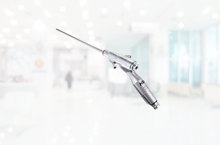
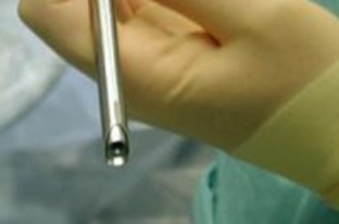
Endoscopic spine surgery is the latest advancement in minimally invasive spine surgery. This procedure uses a endoscope: a device with a small lens at the end and a thin tunnel to pass instruments for removing discs, bone spurs, and tissue.
Endoscopes are commonly used by orthopaedic surgeons in the knee, shoulder, and hip. Recent advancements allow surgeons to use scopes in the spine as well. Patients suffering from compressed nerves are candidates for the procedure.
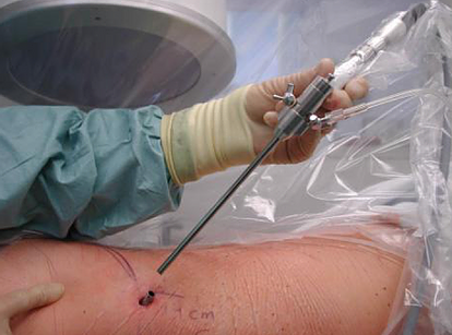
The surgeon first makes a skin incision as small as 7mm. The scope is then placed through this small incision to the disc herniation. The disc herniation is then removed. This incision is smaller than any minimally invasive microdiskectomy, laminectomy, “laser surgery,” or “ultrasonic surgery.”
Potential benefits of endoscopic surgery:
- Possibly avoid fusion surgery
- Small incisions (1cm or less)
- Less pain after surgery
- Lower risk of infection
- Quicker return to normal activities
The procedure is covered by many health insurances.

Lens inserted through a small incision

The Endoscope

The Lens
Surgical Technique Video
This video highlights the basic steps:
- Surgeon targets the disc.
- Muscles are dilated instead of being cut.
- The endoscope is inserted
- The surgery is performed on a video screen
- Through the endoscope, the surgeon can pass instruments to remove disc herniations or bone spurs.
Patients go home shortly after the operation.
Patients receive a printout of their surgical images.


